Feature Story
As published in the UConn Advance, November 3, 2008.
Gift Provides for Integrated Diagnostic and Treatment Suite
By John Sponauer

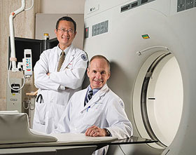
Dr. Bruce Liang, left, director of the Pat and Jim Calhoun Cardiology Center, and Dr. Douglas Fellows, chair of the Department of Diagnostic Imaging and Therapeutics at the Health Center.
Photo by Lanny Nagler
A new major gift will enable the Health Center to offer an integrated imaging and treatment suite.
The suite will help patients move seamlessly from diagnostics to treatment, using the latest technology. It will be the first of its kind in the region.
Torrington natives Carole and Ray Neag made the $3.8 million pledge to upgrade the Health Center’s computed tomography (CT) scanner with a new, more advanced model, and to incorporate new planning and treatment tools into the suite.
The latest pledge complements their 2006 gift to acquire a TomoTherapy cancer treatment system for the Carole and Ray Neag Comprehensive Cancer Center.
The new integrated suite will allow for even more thorough and precise application of TomoTherapy.
The suite will enhance nearly every area of the Health Center’s operations, from conducting research to educating students and treating patients through the Center’s signature programs, such as cancer and cardiology.
“The suite’s functionality for cardiology alone will be leaps and bounds beyond our existing capabilities,” says Dr. Bruce Liang, director of the Pat and Jim Calhoun Cardiology Center. “This will truly be an upgrade to state-of-the-art technology.”
Selective images
Advantages of the new scanner include clearer images, a reduction in
scanning times by about 90 percent, and selective presentation of a
scanned image, allowing a physician to, for example, isolate the
image of a heart without including arteries and vessels that may be
blocking the view.
The suite also offers a CT simulator for treatment planning, and new high dose rate (HDR) brachytherapy, used to treat breast, cervical, uterine, and other cancers at the Neag Comprehensive Cancer Center.
Brachytherapy is a treatment that uses localized, controlled radioactive implants placed internally near cancer cells. Traditionally, brachytherapy required the patient to be on bed rest for several days, with limited exposure to others because of radiation.
HDR is offered on an outpatient basis, with 20-minute treatments, typically repeated a few times.
The addition of the simulator allows more efficient, convenient, and accurate treatment planning to be offered for all cancer patients.
“The addition of HDR dramatically increases our ability to give our cancer patients treatment options,” says Dr. Robert Dowsett of the Division of Radiation Oncology.
“It is becoming the standard of care, and offers major improvements in patient convenience and comfort.”
Ray Neag says the decision to give was driven by the desire to offer state residents the very best care.
“Carole and I take a broad view about the need to serve the people of Connecticut,” he says.
“We feel strongly about the state and its University, and believe that our state’s flagship research university should have the very best, if at all possible.”
Dr. Douglas Fellows, chair of the Department of Diagnostic Imaging and Therapeutics at the Health Center, says the new equipment couldn’t come at a better time in the field’s development.
“As radiology advances, it’s important that we remain on the cutting edge at the state’s flagship teaching hospital,” he says.
“The Neags’ generosity has made a huge difference to UConn and the patients who depend on us every day.”


