

Feature Story
Health Center Today, September 23, 2010
Making Movies Within Cells
By Christine Buckley
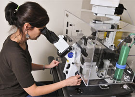
Barbara Mellone, assistant professor of molecular and cell biology, in her lab.
Photo by Jessica Tommaselli
On the second floor of Beach Hall on the Storrs campus in her newly renovated biology lab, Barbara Mellone has a complex set of high-tech cameras. She uses these cameras to snap high-resolution photos – and even make videos – of her research organisms, which can often be elusive. But these cameras aren’t those large digital ones with manual focus and interchangeable lenses that many photographers use.
In fact, they’re small enough to fit into the lens of a microscope.
Mellone, an assistant professor of molecular and cell biology in the College of Liberal Arts and Sciences, studies the process of cell division, in which cells split in two to create new, “daughter” cells. Her goal is to identify the molecular events that lead to successful cell duplication, and these cameras let her and her students watch the action as it happens.
“It gives us a fantastic way to monitor chromosome segregation,” she says. “It’s much easier to see when something’s going wrong.”
Living cells are constantly dividing, as old cells die and new ones take their place. When new cells are needed, the cells replicate their DNA within chromosomes, and using a central region called a centromere, they split the genetic material equally into two new cells.
At least, that’s what’s supposed to happen.
Mellone is interested in what happens when a dividing cell makes a mistake. If the daughter cells are accidentally given too many or too few chromosomes – a condition scientists call aneuploidy – it can lead to dysfunction and cell death. And if those cells happen to be reproductive cells like those that make up sperm and eggs, aneuploidy can lead to birth defects, conditions such as Down syndrome, and even death of the developing embryo. Aneuploidy is also common in tumors and cancer cells, says Mellone.
“You need a robust centromere connection to have good replication,” she says. “In tissue that divides endlessly, like cancer cells, there’s more of a chance that something will go wrong. The cells need a fine balance of what’s necessary to propagate.”
This is where her specialized cameras come in. Using a technique that labels proteins to express different fluorescent colors, Mellone and her students take time-lapse photography of cells dividing under the microscope. The result is a cell division video – a colorful, revealing look at just how each cell part functions over time.
Mellone was recently awarded a four-year, $730,000 grant from the National Science Foundation to study how centromeres assemble themselves in the fruit fly Drosophila. She will use the funds to study the role of CENP-A, an essential protein required for centromeres to exist, which is found in all organisms with cell membranes, including humans.
“CENP-A is a special protein that marks DNA specifically at the centromere,” says Mellone. “Like all centromeric proteins, it is essential for survival. If you don’t have CENP-A, you’re dead.”
Mellone uses her time-lapse photography not only in her research, but also in the classroom and to teach students in her lab. Teaching students about the cell cycle from a textbook can be difficult and confusing, she says, but viewing movies of the cycle can make it much clearer. She recalls showing her videos in a graduate class she taught in her first year at UConn.
“The students were just blown away,” she says. “When you just talk about the different stages, it’s very abstract. It makes so much more sense when you can visualize the process.”
Mellone has used her videos in an interdisciplinary science course for female freshmen and sophomores that she helped develop in collaboration with UConn’s Women in Math, Science, and Engineering (WiMSE) Learning Community.
This summer, she also mentored three local high school students in her laboratory through UConn’s Mentor Connection program. Hailing from Putnam High School, Convent of the Sacred Heart, and Crosby High School, the students were actively engaged in the molecular research in Mellone’s laboratory, including learning how to make these videos and interpret their results.
Although Mellone studies theses processes in fruit flies, the concepts can be extended to inform what we know about cell division in all multicellular organisms. She hopes her studies will help scientists understand how these highly specialized processes have evolved differently in different species.
“It’s becoming more and more obvious that there’s so much more we need to learn about these processes,” she says. “These studies in Drosophila tell us about the mechanisms behind the events, and how they’ve evolved.”